-
Notifications
You must be signed in to change notification settings - Fork 16
MRI_Heights_of_Surfaces_Tools
The tools help to compare the height in the z-dimension of the signals in different channels. It calculates the heights normalized by the maximum height in a reference channel. Only places where the signal is not zero and the reference channel maximal are taken into account. You can find a synthetic example image here: sdata03.tif.
The source code in git-hub can be found here.
To install the tools, drag the link heights_of_surfaces_tools.ijm to the FIJI launcher window and save it under macros/toolsets in the FIJI installation.
Select the "heights of surfaces tools" toolset from the >> button of the ImageJ launcher.
- the first button (the one with an image) open this help page
- the b-button runs the background correction on the input image
- the c-button creates a 2D-surface image in which the pixel values correspond to the max. z-position at a point after the image has been thresholded
- the p-button will display a line-profile plot of the different channels.
- the h-button will calculate the heights of the signals in the different channels.
- the last button (with the gear-icon) will run the batch processing on a folder containing the input images
Options are available for the the creation of the surface image the creation of plots of the heights and the batch-processing. The options dialogs can be opened by a right-click on the c, p and gear-buttons.
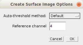
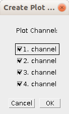
- Auto-threshold method: The method used to automatically determine the threshold for each channel
- Reference channel: The channel that defines the maximal height
- Plot_channel: Select the channels, which will be displayed in the plot
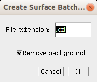
- File extension: The file extension of the input image files
- Remove background: If selected the background correction will be used in the batch processing
The method tries to find background regions and calculates the mean-background intensity per z-slice. The mean value of each z-slice is subtracted from the slice.
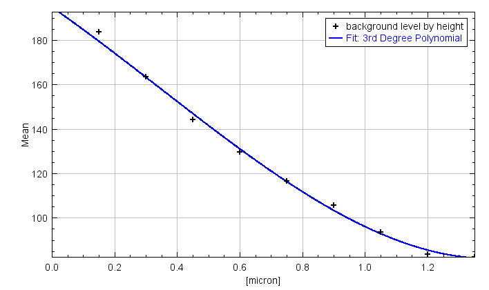
First regions that are clearly background for all z-positions are detected in the channel. The mean intensity per z-position, in the background regions, is measured and a 3rd degree polynomial is fit to the data, in order to smooth it. The resulting mean background intensity is subtracted for each z-position.
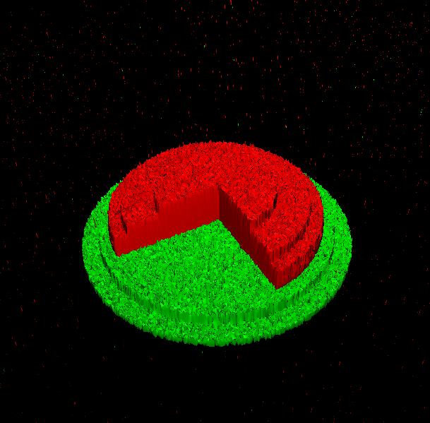
The image is thresholded and a new 2d-image is created, in which each pixel value represents the maximum z-position of the pixel for which the thresholded intensity is not zero.
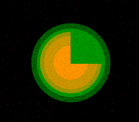
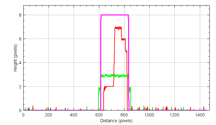
A line is drawn on the image and a line plot is displayed. The the x-axis represents the distance from the start of the line and the y-axis the maximum height of the signal. The result for both channels is displayed in the same plot. If the image already has a line selection when the command is run, the already existing selection is used, otherwise a default selection from the upper-right corner to the lower left corner is used.
The heights of the surfaces are reported in the results table. The columns Mean to Median report the absolute values, while the remaining columns report the values relative to the maximal height in the reference channel.
When you run the batch-processing a first dialog will ask for the path to the folder containing the input images. All images that have the right file extension in the subtree of the input folder will be processed. A second dialog will ask for an output folder. The line plot, a control image of the 2D-surface image, the image with the measured regions as overlays (par default not displayed but accessible via the Overlay-menu in ImageJ) and the final results-table, containing the measurements of all images will be saved into this folder.
28.02.2020 First version
-
El Alaoui, F., Casuso, I., Sanchez-Fuentes, D., Arpin-Andre, C., Rathar, R., Baecker, V., Castro, A., Lorca, T., Viaud, J., Vassilopoulos, S., Carretero-Genevrier, A., Picas, L., 2022. Structural organization and dynamics of FCHo2 docking on membranes. eLife 11, e73156. https://doi.org/10.7554/eLife.73156
-
Sansen, T., Sanchez-Fuentes, D., Rathar, R., Colom-Diego, A., El Alaoui, F., Viaud, J., Macchione, M., de Rossi, S., Matile, S., Gaudin, R., Bäcker, V., Carretero-Genevrier, A. and Picas L. (2020). Mapping Cell Membrane Organization and Dynamics Using Soft Nanoimprint Lithography. ACS Appl. Mater. Interfaces acsami.0c05432.


 Volker Bäcker